Feb. 17, 2021
Competition focuses on microscopy images that help colour-blind researchers

Imagine rolling up to a traffic light and not being certain it’s red. Or going to the grocery store and not being able to see whether the fruit you’re about to grab is ripe.
Now put yourself in the shoes of an academic researcher looking under a microscope and the colours present a barrier to “seeing” the scientific images correctly. If you’re colour-blind, this is your reality.
“Colour-blindness is quite uncommon for females, so I wasn’t identified as colour-blind until university. I just thought I had terrible fashion sense!” jokes Dr. Kamala Patel, PhD, a professor in the Cumming School of Medicine’s (CSM) departments of Physiology and Pharmacology, and Biochemistry and Molecular Biology.
Colour-blindness, or colour vision deficiency, is the decreased ability to see colour or differences in colour. One in 12 men and one in 200 women are colour-blind, more than 95 per cent having red-green colour-blindness.
“As a researcher and microscopist, I have struggled with the red-green colours that are commonly used. I started telling my students that if they use red and green, I wouldn’t be able to see or understand their data,” says Patel, a member of the CSM’s Snyder Institute for Chronic Diseases and associate member of the Hotchkiss Brain Institute.
Advanced, high-resolution microscopy used by University of Calgary researchers is key to observing the natural world and is a mainstay in medical research. The most common colours used in microscopy images are blue, green and red. Shown individually, these can be challenging for a colour-blind person to interpret, but when these images are overlayed, it is nearly impossible. This problem can be avoided by producing images using different colour palettes designed to be accessible to those who are colour-blind.
“The solution is quite simple, it’s just not that well known. Most of the cameras we use for advanced microscopy use greyscale. To make the data accessible, my team now chooses magenta and cyan, both of which are on the colour-blind palette,” Patel says.

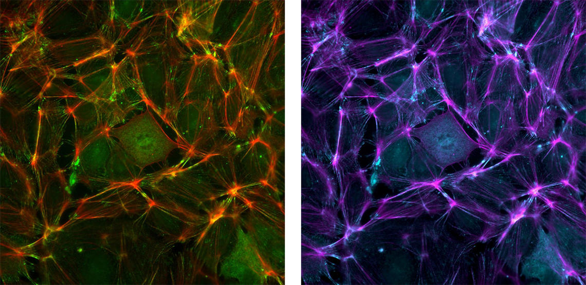
These are images of endothelial cells that line the blood vessels. The first, on the left, uses standard red to label actin and green to label a focal adhesion protein. The second image, on the right, is the same image using magenta and cyan instead.
Kamala Patel
The Striking Image competition has been an annual event since 2010. To raise awareness about colour-blindness, this year’s competition is seeking microscopy images that adhere to the best practices of colour-blind accessibility. The challenge is open to researchers across UCalgary.
“Honestly, most researchers don’t give making their images colour-blind friendly much thought, which is the problem and what we are advocating for,” notes Dr. Aaron Goodarzi, PhD, an associate professor in the Department of Biochemistry and Molecular Biology and member of the CSM’s Arnie Charbonneau Cancer Institute. He is also director of the Charbonneau Microscopy Facility, which is supporting the competition.
Researchers in Calgary formed an alliance of core microscopy facilities involving CSM research institutes, Kinesiology and Alberta Health Services several years ago. They work together, sharing best practices, aligning philosophies for training and service delivery, and generally supporting each other when a need arises.
The contest closes on March 1 at noon and winners will be announced at a virtual ceremony in April.
“I hope this competition raises awareness about how simple steps can both create beautiful images and, more importantly, make images that can be understood by more of the community,” says Patel. “I hope it sparks a conversation about colour-blindness in scientific research, about ensuring equitable and inclusive learning and working, and removing barriers. Even the ones we can’t always see.”
This event is made possible with support from Nikon Canada, Olympus Canada, Quorum Technologies, ThermoFisher, Thorlabs, and Zeiss Canada.
Dr. Kamala Patel, PhD, is a professor in the departments of Physiology and Pharmacology, and Biochemistry and Molecular Biology, and a member of the CSM’s Snyder Institute for Chronic Diseases and associate member of the Hotchkiss Brain Institute. She is the executive director of the Snyder Institute’s Live Cell Imaging Laboratory.
Dr. Aaron Goodarzi, PhD, is an associate professor in the Department of Biochemistry and Molecular Biology and member of the CSM’s Arnie Charbonneau Cancer Institute. He is the director of the Charbonneau Microscopy Facility and holds the Canada Research Chair for Radiation Exposure disease.
